


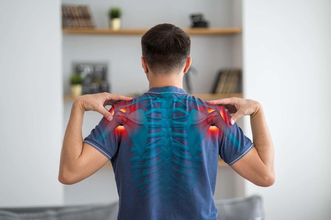
Osteoarthritis of the shoulder is a dystrophic lesion of the cartilage plate that covers the articular surfaces of the joint, which then affects the underlying bones.
About this disease
With this disease, not only the cartilage and subchondral bone are affected. The progressive pathological process also involves the joint capsule and ligamentous apparatus, synovium, muscle and tendon compartments, as well as the subacromial region.
Shoulder arthritis at a certain stage can lead to the development of osteoarthritis. This condition is characterized by the following symptoms: chronic pain, reduced range of motion in the joints, intra-articular brittleness when rotating. Usually, people over 40 years old are subject to this transition.
The main symptoms of shoulder arthritis are pain and limited arm mobility. To verify the diagnosis, informative imaging methods are used - ultrasound and X-ray, computed tomography and magnetic resonance imaging.
According to clinical recommendations, treatment of the disease in the early stages is carried out using conservative methods, and in the later stages, when the cartilage layer is significantly damaged and the patient's ability to self-care is impairedJoint replacement is indicated.
Types of arthritis of the shoulder joint
According to the classification, the following types of shoulder arthritis are distinguished:
- primary joint diseases, in the development of which genetics plays a large role and even the most thorough examination does not allow us to determine the most important cause of the disease;
- Secondary arthritis is a consequence of the impact of adverse factors on the joints (trauma, endocrine diseases, impaired joint anatomy).
Doctors evaluate the rate of progression of the pathological process according to the severity of the disease. The more intense the process, the faster the destruction of articular cartilage and involvement of the underlying bone. Morphologically, the shoulder joint has 6 levels of osteoarthritis:
- first degree - the cartilage matrix becomes swollen and disintegrates, but the integrity of the surface area of the cartilage remains intact;
- second degree – the cells of the cartilage tissue located in the deep layers are affected, the surface plate of the cartilage is damaged;
- third degree - longitudinal cracks appear on the cartilage plate;
- fourth degree - the surface area of the cartilage plate gradually peels off, erosive defects are formed and cystic cavities appear in the underlying bone;
- fifth degree - at this stage the underlying bone is exposed;
- sixth degree - the subchondral area thickens significantly, the cysts become more pronounced and marginal bone growth appears.
Symptoms of shoulder osteoarthritis
The main clinical signs of shoulder arthritis are pain and stiffness to the point of complete loss of mobility as well as joint deformity.
Outstanding features of pain due to joint deformity are:
- appears when bending, extension, or rotation begins;
- increases during physical activity;
- nocturnal nature due to stagnation of venous blood in the visceral channels;
- the presence of blockade - sudden jamming in the joint due to the separation of fragments of bone and cartilage separating between the articular surfaces;
- depends on the weather - the pain increases when the weather changes (in humid and cold climates, the pain becomes more intense).
Arthritis is a chronic disease. In the early stages of the disease, pain appears cyclically (at a time when the disease worsens). The rate of progression of the pathology is determined by the timeliness of initiating treatment and the adequacy of lifestyle adjustment. Shoulder pain becomes chronic if it lasts 6 months or more. The change from acute to chronic pain indicates progression of the pathological process.
Causes of shoulder arthritis
Causes of shoulder osteoarthritis are divided into 2 groups:
- modifiable - repairable;
- unmodifiable - cannot influence their actions.
Non-modifiable factors that may increase the risk of developing joint changes in the shoulder joint include:
- gender - by the age of 50, women are less susceptible to diseases than men, after about 50 years, the incidence of pathology in both sexes becomes approximately the same;
- human age - the older the patient, the higher the risk (and from about 30 years of age in cartilage tissue, the degeneration process occurs faster than the regeneration process, creating a premise for the disease to develop);
- congenital abnormalities of the shoulder structure - increased hypermobility (hypermobility), connective tissue dysplasia (usually, articular cartilage is represented by type 2 collagen fibers, with dysplasia, replacement occursby less durable collagen), joint instability;
- genetic characteristics - genetically determined predominance of type 2 collagen, polymorphism of interleukin-1 and interleukin-2 genes.
Modifiable risk factors for right or left shoulder deformity are:
- joint injury;
- excessive physical activity (strength sports and martial arts, including weightlifting);
- obesity – for shoulder arthritis, the important factor is not the increase in mechanical load, but the metabolic changes that occur in connective tissue, incl. chronic inflammation that accompanies obesity;
- weakness of the Corset muscle of the shoulder joint, especially in people who perform activities that require manual meticulousness (jewelers, dentists, secretaries, writers);
- lack of vitamin D, which actively participates in maintaining the health of the musculoskeletal system;
- a diet low in vitamin C, an important link in the body's calcium-phosphorus metabolism;
- Hormonal imbalance – thyroid disease, diabetes, etc. v. ;
- smoking - both active and passive.
In shoulder arthropathy, the main targets of the pathological process are the articular cartilage, subchondral bone and synovial membrane. In affected cartilage, proteoglycan synthesis is reduced, fragmentation and cracking of the plate is observed, exposing the underlying bone. Increased non-physiological load on the bone leads to its compaction, the appearance of cysts and bone spurs (marginal growth).
Diagnose
Examination of a patient with shoulder pain should begin with an X-ray. It is important to scan multiple views to check joint details. Images can be captured in direct projection mode, in internal and external rotation positions. To evaluate the soft tissue formation of joints, especially in the early stages of joint disease, joint ultrasound is the most useful information. If the diagnosis remains unclear, magnetic resonance imaging/computed tomography arthrography is recommended. At the next stage, the preservation of joint functions is evaluated.
Expert opinion
All morphological forms of the joints are involved in the pathological process. The main symptom of osteoarthritis is pain in the joint area, not only due to synovitis but also due to bone damage (osteitis, periostitis), involving soft tissues around the joint (tendinitis, tenosynovitis). , myalgia, enthesopathy, capsular dilatation), meniscal degeneration, and involvement of the sensory nervous system (eg, irritation of nerve trunks by large osteophytes). Therefore, the sooner treatment begins, incl. Lifestyle modifications will make pain control more effective.
Treatment
At the initial stage of the pathological process, treatment of shoulder osteoarthritis is carried out using conservative methods, and with severe degeneration of articular cartilage, surgical intervention (endoscopy) is required.
Conservative treatment
During the exacerbation phase of the process, the first priority is pain relief. Nonsteroidal anti-inflammatory drugs are often used to relieve pain. They can be applied topically (in the form of creams and ointments), injected into the joint cavity or used systemically (tablets, intramuscular injections). In some patients, the pain may be so severe that a short course of corticosteroid medication may be used for pain relief.
Intra-articular injections of hyaluronic acid or plasma, incl. enriched with platelets, can have a stimulating effect on the cartilage plate and promote its renewal (this treatment is considered pathogenic). These injections help accelerate the synthesis of collagen and elastic fibers that form the foundation of cartilage. As a result, the structure of the cartilage layer and synovial membrane is improved, helping to increase the uniformity of joint surfaces. These injections into the joint help optimize the production of synovial fluid, which not only absorbs shock and moisturizes the cartilage, but also improves metabolism in cartilage cells, increasing their internal potential.
After the acute process subsides, physiotherapeutic rehabilitation methods (pulsed current, ultrasound and laser treatment) can be used as part of complex treatment. These procedures have a complex positive effect on joint structures.
Surgery
This surgery is indicated for significant destruction of the cartilage plate, accompanied by persistent pain and joint dysfunction, leading to the inability to self-care and perform professional tasks. A modern method of surgical intervention for shoulder arthritis is arthroscopic implantation. At SM-Clinic, surgery is performed in strict accordance with the method of using the latest generation of prosthetics. This is the key to achieving the best treatment results.
Prevent shoulder osteoarthritis
The main prevention of shoulder osteoarthritis is aimed at maintaining optimal metabolism in the osteochondral cavity. For this it is recommended:
- maintain normal body weight;
- Adequate compensation of endocrine disorders in the body (needs consultation and dynamic monitoring by an endocrinologist);
- Dosage strengthens the muscles of the shoulder girdle;
- Warm up regularly if your professional activity involves performing similar shoulder movements.
To prevent the progression of shoulder arthritis that has developed, the following recommendations are important:
- Avoid lifting heavy objects, incl. dumbbell push-ups;
- conduct repeated therapeutic massage courses;
- Regularly participate in health-enhancing exercise (under the supervision of a physiotherapist).
Rehabilitation
After endoscopy, a layer of plaster is applied, which provides the necessary degree of fixation. After removing the cast, the period of restoring functional activity of the joint begins. To do this, it is recommended to take massage therapy, physiotherapy and health-promoting fitness courses under the supervision of a physiotherapist.
Questions and answers
Which doctor treats shoulder osteoarthritis?
Diagnosis and treatment of the disease are performed by an orthopedic traumatologist.
Representatives of which professions most often suffer from shoulder arthritis?
Athletes participating in volleyball, tennis, basketball, shot put and excavator sports are at the highest risk of destruction due to degeneration and dystrophy of the cartilage of the shoulder joint.
Is shoulder pain a sign of arthritis?
Indeed, pain is the leading sign of arthritis. However, pain can also be a symptom of other diseases - adhesive capsulitis, osteoarthritis, rotator cuff damage, etc. v. A qualified orthopedic traumatologist will help you make an accurate diagnosis and choose a treatment method.